

PELCO® NiOx Test Specimen for Analytical Electron Microscopy (AEM)
Inexpensive characterization of instrument performance
|
The NiOx Test Specimen has been developed for advanced analytical TEM's equipped with X-Ray Micro Analysis Systems (EDX) and/or Electron Energy Loss Spectrometers (EELS) to characterize instrument performance. It consists of a 55nm layer of NiOx on a 25nm carbonsupport film on a 300 mesh Mo TEM grid. The NiOx Test Specimen is valuable in at least three ways:
The test specimen provides an Mo signal to measure stray X-rays and electrons present in the TEM column. The NiOx film contains a known amount of a light element (oxygen) and is used for evaluation the EDX detector response to low energy X-rays. In EELS, the oxygen and nickel ionization edges allow energy-axis calibration and elemental-ratio measurements to be performed. It is possible to make a direct comparison of elemental analyses obtained by EDX and EELS for a TEM equipped with both systems. |
||
 |
 |
|
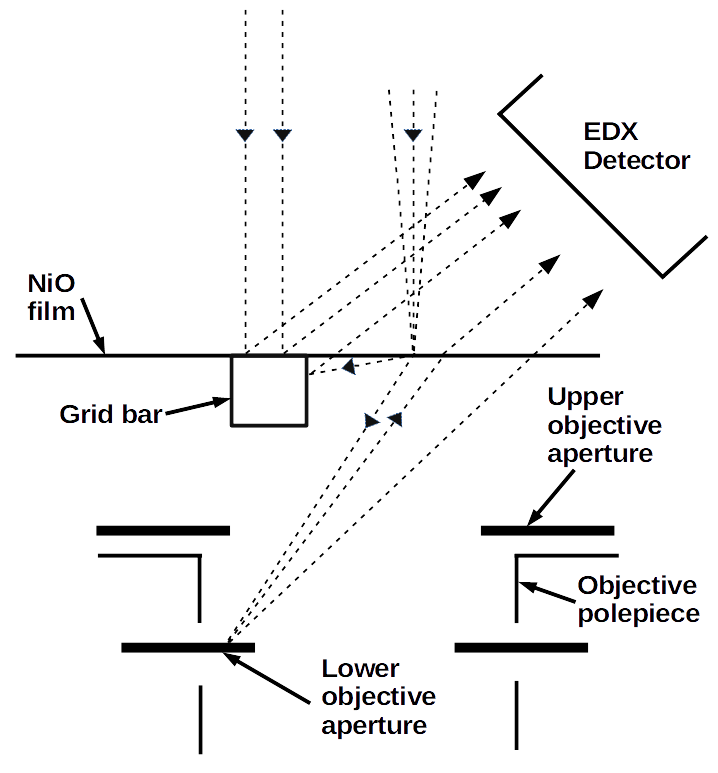
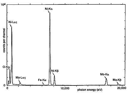
| Spurious X-rays (dashed trajectories) created when scattered electrons strike a grid bar (not to scale) or and objective aperture, or by fluorescence of the grid by column X-rays.; | Typical EDX spectrum for a 55nm specimen of NiO on a 300-mesh Mo grid, coated with 25nm C, with the 200 keV incident beam near the center of a grid square. The small Fe Ka: peak is generated by the microscope column (perhaps objective pole-pieces). (reprinted with permission) |
|
 Since the nickel oxide film is a fine-grained polycrystal, it is well suited to the calibration of TEM camera length and the correction of intermediate-lens astigmatism. Each specimen is packaged in an aluminum storage container #650-10 with instructions for the measurement of camera length, evaluating the amount of stray radiation in the TEM column and its predominant character (X-rays or electrons), for estimating the collection solid angle of the EDX detector and for determining the extent of ice or hydrocarbon contamination on the detector. 1. Egerton RF, Cheng SC, (1994) Characterization of an analytical electron microscope with a NiO test specimen. Ultramicroscopy, 55 43-54. |
||